3D Imaging, based on X-ray and neutron technologies, is a non-destructive technique that provides 3D images of the internal structure of materials and components.
 Imaging at DTU specialises in the 3D Imaging technique called computed tomography (CT) with X-ray and neutrons.
X-ray computed tomography (X-ray CT) was originally developed for medical imaging in the 1970's and has evolved to become a widespread technique for industrial applications, where it is used to produce 3D images of the inner structure of materials without having to cut or slice the materials.
X-ray and neutron CT consists in irradiating a material with an X-ray or neutron beam, while the material sample rotates 360°. A detector captures thousand of images based on the material's ability to absorb the X-ray or neutron beam. The data set captured by the detector is then analyzed thank to sophisticated software and powerful computers, which allow the reconstruction of 3D images of the material. The 3D reconstruction can also be sliced to show the internal structure layer by layer, in analogy to pealing an onion. This is illustrated by the short movie above.
X-ray and neutron CT: complementary technologies
X-ray and neutron technologies are complementary and can be used to study different kinds of materials or different properties in materials. Said very basically, neutrons are absorbed by light elements (i.e. with low atomic numbers) such as hydrogen, water, carbon etc., whereas X-ray are absorbed by heavy elements (i.e. with high atomic numbers) like metals for example.
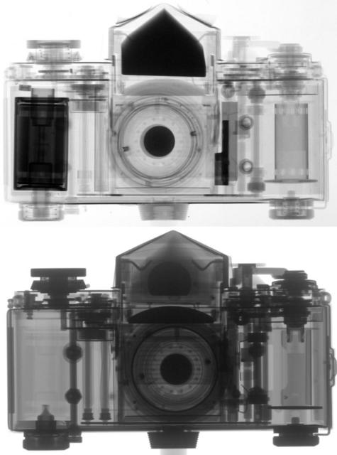 These two pictures illustrate the complementarity of X-ray and neutron imaging.
A neutron beam (top) easily penetrates the outer metal cabinet of the camera
but is instead absorbed by the inner plastic parts, which appear dark.
On the contrary the X-ray beam (bottom) is absorbed primarily by the metal parts.
3D Imaging at large scale facilities
Because of a very broad spectrum of applications - both in fundamental research and for applied industrial projects - 3D imaging is one of the fastest expanding fields at large scale facilities all around the world. The most advanced large-scale facilities in Europe are ESRF and ILL in France, and SLS and SINQ in Switzerland.
Moreover, two new facilities are currently under construction in Lund, Sweden: European Spallation Source (ESS), the world's best neutron source, and the MAX-IV synchrotron, a unique X-ray source. In combination these two facilities will be the largest microscope in the world and provide unique opportunities for investigating all kinds of materials.